")
")
Latest News
Social Media
| Â | |||
| Â |
 |
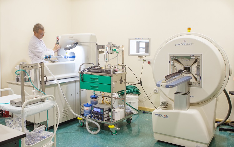
Small animal PET and CT
The CNA small-animal PET scanner was made by Philips (model Mosaic). The detection system is based on 14,456 GSO crystals (Gd2SiO5:Ce, gadolinium orthosilicate activated with cerium) with dimensions of 2x2x10 mm3 and is arranged in 52 rings of 278 crystals each. The GSO crystals are glued to a continuous light guide with a thickness of 1.2 mm and 0.5 mm deep slots. The crystals are read out by a hexagonal array of 288 photo multiplier tubes (PMTs) with a diameter of 19 mm each. The scanner operates exclusively in 3D mode and the spatial resolution is 2.7 mm at the centre. The unit also has capability for housing up to thirty animals in environmentally controlled rooms.
Â
In October 2008, a new preclinical microCT system (made by Bioscan, model NanoCT) was installed by Philips Medical Systems. The NanoCT system is a helical CT scanner which operates at 65 kV maximum voltage, obtaining images with a spatial resolution higher than 200 ÎĽm. The scanner has an axial field of view (FOV) of 270 mm and a transaxial FOV of 76 mm. This unit will be used to obtain computed tomography (CT) images with X-Rays from small animals and objects of technological or archaeological interest.
Â
The system is equipped with exploration beds that are fully compatible with the Mosaic PET scanner. Sequential image acquisition with both techniques (PET/CT) in experimental animals is thus possible and a unique multimodality PET/CT image is finally obtained. This single set of images combines PET metabolic and CT morphologic information.
Â
The laboratory has several programs for the evaluation and analysis of CT, PET images and other modalities as MRI: imalytics, Invivoquant 1.43 and PMOD 3.6 for which has the maintenance contracts which allow to update always to the newest version of the program. The most used one, PMOD, has from 2014 the following modules: VIEW (basic operation with the images, batch image processing), KINETIC (kinetic modelling of the radiotracers), PXMOD (pixel by pixel parametric mapping of kinetic models), FUSION and FUSEIT (for fusion/overlay of the images of different modalities, normalization to brain templates of human, rat, mouse or macaque [cynomologus monkey]), SEGMENT (for image segmentation according to the pixel values), ALZHEIMER (evaluation of the patient with clinical signs of Alzheimer´s disease based on FDG-PET scan; this is not a clinical tool), NEURO (semiautomatic delineation of the regions of interest of the brain according to nuclear anatomy in PET or MRI images).
Â
 Â
  Â